Beyond Lab Consulting

  • Home
  • About BLC
  • Services
    • Laboratory Survey Management
    • New Lab Set Up with Turn-key Operation
    • Monthly Management
  • Industry News
    • BLC Tips
    • Lab Testing and Common Diseases
  • Testimonials
  • BLC FAQ’s
  • Careers
  • Contact Us
  • Call Us: 631-500-1550

Liver Levels: The Low-Down on Liver Function Lab Tests

September 29, 2018 By Larry Fox

Image courtesy of the National Institutes of Health

By Cat Troiano

According to the American Liver Foundation, nearly 30,000 adults are diagnosed with liver cancer each year, and the number of mortalities exceeds 20,000 annually. Additional diseases threaten the liver, including some that can be just as deadly and some that can be prevented. Evaluating liver function is a key first step toward detecting liver disease at an early stage.

Liver at Large

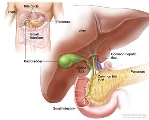
Weighing in at roughly three pounds, the liver, which is situated on the right side of the abdomen, is one of the largest organs of the human body, ranking as the second-largest organ after skin. The liver is unique in that when it sustains damage, it has the ability to regenerate as long as at least 25 percent of its tissue remains healthy. Technically a gland that is classified as part of the digestive system, the liver carries a lot of responsibilities. Functions of the liver include:

• The liver produces bile, a substance that aids in digestion and is made up of bilirubin, electrolytes, cholesterol and water.
• The liver produces albumin, which is a protein found in blood serum.
• The liver breaks down and metabolizes proteins.
• The bile that is produced in the liver metabolizes fats.
• The liver metabolizes carbohydrates to maintain blood glucose levels.
• The liver stores carbohydrates as glycogen, which can be called on by the body for use when a burst of energy is needed.
• The liver also stores a supply of vitamins, including A, B12, D, E and K.
• The liver also stores ferritin, a form of iron that comes from hemoglobin and is utilized in the production of new red blood cells.
• The liver filters hormones as well as foreign substances, such as drugs and alcohol, from the blood.
• The liver plays a role in the body’s immune function.

With so many vital roles that affect overall health, monitoring the liver’s ability to carry out these tasks efficiently is an important step in diagnosing diseases that can hinder liver function.

Diseases of the Liver

There are several diseases that affect the liver. Some examples of liver diseases include:

• Cirrhosis – a disease in which scar tissue overtakes healthy liver tissue. This process, called fibrosis, has a number of causes, including excessive alcohol use, toxins and hepatitis. Alcoholic liver disease is the leading worldwide cause of cirrhosis of the liver.
• Hepatitis – a liver infection in which the liver becomes inflamed. Viruses, toxins and autoimmune response can cause hepatitis.
• Fatty liver disease – a condition in which fat accumulates in the liver’s cells. While alcoholism is a frequent culprit, obesity can also result in fatty liver disease.
• Primary sclerosing cholangitis (PSC) – an incurable disease characterized by inflammation of the bile ducts. The cause of PSC remains unknown.
• Gilbert’s syndrome – a non-threatening condition in which bilirubin is not fully broken down, resulting in mild jaundice.
• Fascioliasis – a parasitic infection that is caused by liver flukes.
• Liver cancer – the sixth most commonly diagnosed form of cancer

Symptoms of liver disease include jaundice, light-colored stools, dark-colored urine, appetite loss, nausea, diarrhea, fatigue, unexplained changes in weight and abdominal swelling or discomfort, but the liver may sustain extensive damage before symptoms present. Early detection through routine laboratory screening is valuable so that treatment protocols can be initiated to restore the liver’s form and function when possible.

Liver Tests and Values

A comprehensive metabolic profile is an effective routine laboratory test for analyzing a patient’s liver function. There are six results on a comprehensive metabolic profile that represent liver function.

Alanine amino transferase (ALT, or SGPT) is an enzyme that aids in metabolizing protein and converting food into energy molecules. ALT is found primarily in a healthy liver, with only low levels detected in the blood. When the liver sustains damage, however, the damaged cells release ALT into the bloodstream. An abnormally high level of ALT in the blood is indicative of liver disease.

Aspartate amino transferase (AST, or SGOT) is an enzyme that metabolizes the amino acid alanine. AST is normally found in the liver, as well as in the heart and muscles. When the liver, heart or muscles sustain damage, elevated amounts of AST are released into the bloodstream. While a high ALT level is evidence of liver disease, a normal ALT level combined with a high AST level can be indicative of a heart condition or muscle damage.

Alkaline phosphatase (ALP) is an enzyme that is found in the liver, bile ducts and bone. Damage to any of these structures causes an elevation in the ALP level in the bloodstream. High ALP levels may indicate liver disease, a bile duct obstruction or a bone disease.

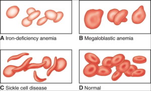
Bilirubin is a waste product that is produced when red blood cells break down at the end of their typical 120-day lifespan. Bilirubin then circulates through the liver, where it is filtered from the blood to exit the body through stool. Liver damage or disease as well as certain types of anemia can result in an elevated bilirubin level. Jaundice is a visible sign in a patient who has a very high bilirubin level.

Albumin is a protein that is produced in the liver, and it accounts for roughly 60 percent of total protein found in the blood. Albumin is essential for nourishing the body’s tissues as it aids in transporting vitamins, minerals and hormones throughout the body. When the liver is damaged, a low albumin level in the blood results.

Total protein, or albumin/globulin(A/G) ratio, represents the combined total of albumin and globulin, which accounts for the remaining 40 percent of protein that is found in the blood. Proteins are the building blocks for all cells and tissues through the body. In a patient with severe liver disease, the level of either albumin or globulin is low.

The normal ranges for liver values are:

ALT: 7 to 55 U/L
ALP: 45 to 115 U/L
AST: 8 to 48 U/L
Bilirubin: 0.1 to 1.2 mg/dL
Albumin: 3.5 to 5.0 g/dL
Total Protein: 6.3 to 7.9 f/dL

Other Liver Function Tests

When any of the aforementioned liver function values yield abnormal results on the comprehensive metabolic profile, additional tests may be ordered to aid in diagnosis. Such laboratory tests include:

Gamma-glutamyl transferase (GGT) is an enzyme that is present in the highest concentrations within the liver. While GGT levels increase as a result of liver damage incurred by any of the liver diseases, it is the first liver enzyme to become elevated when bile duct obstruction occurs. Therefore, the GGT test is useful in determining if an elevated ALP level is due to a blocked bile duct. It can also help to rule out bone disease as a cause of the elevated ALP level.

The normal reference range for GGT is 9 to 48 U/L.

Lactate dehydrogenase (LD, or LDH) is an enzyme that is found in nearly all cells throughout the body. Its highest concentrations are found in those of the liver, kidneys, heart, muscles and lungs, making the result of this test nonspecific when performed alone. When an LD is performed to compare the result against those of the comprehensive metabolic profile, it can be helpful in pinpointing the organ that has sustained tissue damage. A high LD is indicative of tissue damage.

The normal reference range for LD is 122 to 222 U/L.

Prothrombin time (PT) is a measurement of how long the blood takes to clot. Prothrombin is a protein that aids in blood coagulation. Since prothrombin is produced in the liver, the level of prothrombin that is produced can decrease if the liver is damaged or diseased, resulting in a longer blood clotting time.

The normal reference range for patients who are not taking blood thinning medications is 9.5 to 13.8 seconds.

Alpha-feto protein (AFP) is a protein that results when liver cells regenerate. Chronic liver diseases can result in ongoing elevated AFP levels in the blood. The AFP test is also a valuable tumor marker test in that very high levels of AFP are indicative of hepatocellular carcinoma and hepatoblastoma, which are two forms of liver cancer.

The normal reference range for AFP is 0 to 15 IU/ml.

Additional specific laboratory tests, such as the acute hepatitis panel and the autoimmune antibody test, can be ordered to screen for hepatitis A, B and C or autoimmune hepatitis. Diagnostic imaging tests, such as ultrasound or CT scan, may be ordered to visually reveal liver damage, structural abnormalities or masses. A liver biopsy and tissue pathology may be ordered when liver cancer is suspected.

Who Should Be Tested?

While a comprehensive metabolic panel is typically ordered as part of a routine health examination, physicians should also order the screening and/or additional liver tests in patients who are at risk for liver disease. Such situations include:

• Patients who present with the aforementioned symptoms of liver disease
• Patients who are alcoholics
• Patients with family histories of liver disease
• Patients who are overweight or obese
• Patients who may have been exposed to any of the hepatitis viruses

Some herbal remedies and a number of medications, including acetaminophen, non-steroidal anti-inflammatory drugs, statins, hormonal drugs, some antibiotic drugs and antihypertensive drugs as well as certain medications used to treat gastroesophageal reflux, diabetes, gout, allergies, fungal infections, cancer and seizures, can have adverse effects on the liver. Patients who take any such medications should be tested periodically to monitor liver health.

Treatment for liver disease depends on the specific condition that is diagnosed. Some forms of liver disease can be treated with changes in lifestyle habits, such as losing weight or ceasing alcohol consumption, while more severe forms of liver disease may progress to the point of requiring a liver transplant. Adopting healthy lifestyle habits and undergoing periodic routine laboratory tests the enable early detection offer patients the best chance for preserving liver function.


Filed Under: cathy

PSA: What You Need to Know About Prostate Cancer Screening

September 1, 2018 By Larry Fox

By Cat Troiano

According to the American Cancer Society, one out of nine men is diagnosed with prostate cancer, and it is the third leading cause of cancer-related death in men. The present survival rate, in which one out of 41 men succumb to the illness, is higher than the alarming rate of diagnosis, thanks to screening tests.

Risk Factors and Testing Guidelines

Cases of prostate cancer are often detected before any symptoms present. Men who are experiencing changes in urination habits or in reproductive function should consult with their physician, who will order a prostate-specific antigen test and a digital rectal examination at once to screen for prostate cancer. In the absence of these signs, prostate cancer screening recommendations are made based upon each patient’s age and risk factors.

•  Men who are 50 years of age or older and have a remaining life expectancy of at least 10 years should undergo               prostate cancer screening.
• Prostate cancer screening should begin at 45 years of age in men who have elevated risks of developing the illness.

Such risks include:
o Being African-American
o Having a father, son or brother who was diagnosed with prostate cancer before the age of 65
• Men who have a family history of two or more direct relatives who were diagnosed with prostate cancer before the age of 65 should undergo prostate cancer screening from the age of 40.

Prostate cancer is a slow-growing cancer, and thus screening is not typically advised for men who do not have an expected remaining lifespan of at least 10 years. In these cases, the men who happen to have prostate cancer will more likely die with prostate cancer rather than from prostate cancer.

What Is PSA?

The prostate is a male reproductive gland that is located underneath the bladder and in between the seminal vesicle and the rectum, and it surrounds the uppermost portion of the urethra. The prostate’s role is to produce prostate fluid, which combines with semen, and to expel semen into the urethra during ejaculation. Prostate-specific antigen (PSA) is a protein enzyme that breaks down the molecular weight of proteins in semen, rendering semen a more fluidlike form. PSA is produced by both healthy and cancerous cells of the prostate gland, and while much of it is released into semen, some is also released into the bloodstream. Men who have prostate cancer will have a significantly higher concentration of PSA in their blood.

Using PSA as a Tumor Marker

The PSA test, which is performed on a blood serum sample, is used as a tumor marker screening method. The most commonly ordered laboratory test for prostate cancer screening is the total PSA test, which provides results that represent the sum of two forms of PSA in the blood. The two forms are known as free PSA and complexed PSA. Free PSA is that which is not bound to other proteins in the blood, a presentation that is more typical from benign, or noncancerous, prostate cells. Complexed PSA, or cPSA, is that which binds with other proteins and is a more typical result from malignant prostate cells. If the total PSA test result is slightly higher than the normal reference range, then a free PSA test may be ordered to help guide physicians in the decision of whether or not to pursue the recommendation of a prostate biopsy. If the free PSA level is high, then the high total PSA level may be the result of a benign condition as opposed to prostate cancer.

The PSA Test Result

The general normal reference range for a PSA test is less than 4 ng/mL in men who are less than 60 years of age.

As long as the result is less than 2.5 ng/mL, the American Cancer Society recommends repeated screenings every two years. If the result is 2.5 ng/mL or greater, then screenings should be ordered annually. The upper normal reference range limits for PSA levels increase in men beyond 60 years of age. The reference ranges for older age groups are:

• 4.5 ng/mL or less for men aged 60 to 69
• 6.5 ng/mL or less for men aged 70 to 79
• 7.2 ng/mL or less for men aged 80 or older

A digital rectal examination (DRE) to palpate the prostate for any lumps, enlargement or other physical abnormalities is performed as part of a routine physical examination. Blood should be drawn before performing a DRE since this can temporarily elevate the PSA level in some patients.

There are other noncancerous causes of an elevated PSA result. Some of these conditions include:

• Benign prostatic hyperplasia, more commonly referred to as an enlarged prostate, is a condition in which the prostate cell production and growth accelerates. The result is a larger prostate that can compress the urethra, which can potentially cause partial or complete urinary blockage. Benign prostatic hyperplasia is part of the aging process and affects most men as they age.
• Prostatitis is an infection of the prostate which is often bacterial in nature. Unlike benign prostatic hyperplasia, prostatitis can affect men of any age, but it is more commonly diagnosed in those who are less than 50 years of age.
• Urinary catheterization or any medical procedure that can cause irritation or inflammation of the prostate and surrounding structures can lead to an elevated PSA level.
• Ejaculation within 24 hours prior to testing can cause a slight elevation in PSA, which may raise a red flag in someone whose normal PSA level has been borderline on previous screenings.
• Aging raises the normal limits of the PSA level, as noted above.
• Certain chemotherapy drugs can alter the PSA level.

If a PSA test result is higher than the accepted normal range, further diagnostic testing will be ordered to rule out cancer as the cause. Such tests typically include a trans-rectal ultrasound and prostate biopsy. If the pathology report confirms the presence of cancerous cells, the PSA test results can be used in conjunction with the histopathology results as part of the cancer staging process.

Prostate Cancer Treatment Options

Once a patient is diagnosed with prostate cancer, treatment options should be discussed with an oncologist. Some options that are available include:

• Prostatectomy, which is surgical removal of the prostate
• Brachytherapy, or internal radiation therapy, such as the implantation of radioactive seeds
• External beam radiation therapy, such as CyberKnife® radiation
• Cryotherapy, or cryoablation, in which prostate cancer cells are destroyed by freezing
• Hormone therapy to lower the body’s level of androgen, which is a male hormone that stimulates the growth of prostate cancer cells
• Chemotherapy

When discussing treatment options, factors such as age, expected lifespan, the stage of the cancer, the presence of other health conditions and patient concerns regarding potential side effects of treatment must all be considered. Once a treatment protocol has begun, PSA tests can serve to monitor the efficacy and progress of the treatment. Decreasing levels of PSA indicate that the treatment is working.

All male patients should have a prostate cancer risk assessment discussion with their primary care physicians at age 40, and PSA testing should then be ordered in accordance with screening guidelines. PSA testing should also be ordered for all male patients who present with any signs of symptoms of possible prostate problems. Four out of five cases of prostate cancer are diagnosed during the early stage, and when treatment is promptly sought, the 15-year relative survival rate is 96 percent.

Filed Under: cathy

Good for Mom and for Baby Too: What Prenatal Tests Expectant Mothers Should Expect

August 4, 2018 By Larry Fox

By Cat Troiano

Pregnancy is an exciting time for moms-to-be as they anticipate the birth of their baby, and they rely on their physicians for guidance through each trimester. In addition to proper nutrition, stress reduction, plenty of sleep and safe activity levels, periodic checkups are essential in achieving the healthiest possible status for both the mother and for her developing fetus. Prenatal laboratory tests are an important part of these appointments so that any potential health issues can be addressed to ensure a safe pregnancy and uncomplicated delivery.

Types of Prenatal Tests

From pregnancy confirmation through delivery, expectant mothers undergo various laboratory tests to monitor their health as their bodies undergo hormonal shifts and metabolic changes to accommodate the growth and development of their fetuses. The types of prenatal tests that may be ordered include:

• Routine tests
• Selective tests
• Genetic tests

While some tests are ordered for every woman who is pregnant, others are only performed under certain conditions.

Confirming Pregnancy

Human chorionic gonadotropin (hCG) is a hormone that is produced by the cells of the placenta once conception takes place, and the level continues to increase every two to three days during the first 8 to 11 weeks of pregnancy.

A qualitative hCG test evaluates the presence of hCG in a blood or urine sample to confirm a patient’s pregnancy status:

• An hCG level that is lower than 5 mIU/ml is a negative result for pregnancy.
• An hCG level that is between 6 and 24 mIU/ml is inconclusive, and the test will need to be repeated to determine if the level increases.
• An hCG level that exceeds 25 mIU/ml is a positive result for pregnancy.

A quantitative hCG test is performed on a blood sample for monitoring purposes to ensure that the level of hCG is increasing appropriately during the first trimester. A low hCG level is not always indicative of a problem, and plenty of women with low hCG levels have given birth perfectly healthy babies. However, if the level does not increase, problems such as etopic pregnancy and miscarriage must be ruled out. Conversely, a higher than normal hCG level must also be investigated to rule out possible causes, such as a multiple pregnancy.

Routine Tests for All

Once the qualitative hCG test confirms a patient’s pregnancy status, routine laboratory screening tests are ordered for all expectant women.

A complete blood count (CBC) is performed during the first and third trimesters. This blood test, which evaluates the red blood cells, hemoglobin, white blood cells and platelets, enables the detection of the following conditions:

• Anemia, which can occur during pregnancy when the mother isn’t taking in adequate amounts of iron, folate or vitamin B12, posing the risk of delivering a premature or low birth-weight baby
• Infections, which can be passed along to the fetus
• Gestational thrombocytopenia, which is a condition that can develop during mid-to-late pregnancy and results in a low platelet count, reducing the blood’s ability to clot and increasing the risk of hemorrhaging

A urinalysis is performed at each prenatal appointment throughout the pregnancy. This urine screening enables the detection of these conditions:

• Gestational diabetes, which is a form of diabetes that can develop during pregnancy if hormonal changes result in high glucose levels and ketones in the urine
• Preeclampsia, which is a potentially fatal condition that can arise during pregnancy and is characterized by high blood pressure and high protein levels in the urine, putting the mother at risk for seizures, stroke, heart failure or coma and putting the fetus at risk for being stillborn

A urine bacterial culture may also be performed to detect bladder and urinary tract infections.

Blood typing and antibody screening is performed during the first trimester and repeated during the 28th or 29th week of pregnancy to verify that the mother’s and fetus’ blood types are compatible and that there are no Rh factor antibodies present.

A group B streptococcus (GBS) bacteria culture is performed during the 35th to 37th week of pregnancy. A baby can contract GBS during birth, which can result in the newborn developing meningitis or pneumonia.

If any abnormalities are discovered in these test results, additional tests may be ordered. Diligent monitoring, combined with appropriate treatment, is imperative in managing the condition. For example, if the CBC results show a hemoglobin count that is lower than the normal range of 12 to 15.5 grams per deciliter, additional tests, such as the ferritin serum test or the serum folate test, can determine if the anemic state is the result of an iron or folate deficiency, respectively. Once a deficiency is confirmed, appropriate supplementation can then be prescribed to correct the deficiency.

Other tests that are recommended by the American College of Obstetrics and Gynecology for all expectant mothers include screenings for the following diseases, which can result in complications or be passed to the fetus:

• Human immunodeficiency virus
• Human papilloma virus
• Rubella, or German measles immunity
• Varicella zoster virus (chicken pox) immunity
• Hepatitis B
• Chlamydia
• Syphilis

Mothers who have histories of certain medical conditions or who carry other risk factors may need to undergo additional tests.

Selective Tests for Mothers at Risk

Just as an expectant mother who has been managing hypertension should expect diligent blood pressure monitoring throughout her pregnancy to avert the development of preeclampsia, mothers with other health conditions must be equally diligent in staying on top of their health status. Some examples of laboratory screenings that are ordered in such cases include:

• The glucose challenge and/or glucose tolerance test, for those with pre-existing diabetes, those who have a prior history of gestational diabetes or those whose urinalysis results include elevated glucose levels and ketones
• TSH, for those with a history of a thyroid condition, such as hypothyroidism
• Zika virus test, for expectant mothers who have recently traveled to areas where this mosquito-borne illness is prevalent
• Tuberculosis test, for those who are HIV positive or who reside with someone who has tuberculosis

To minimize the chances of complications during pregnancy, labor and delivery, attentive monitoring and testing are recommended for all expectant mothers who carry higher risks. Some of these risks include:

• Obesity
• Pregnancy that was achieved with the help of fertility drugs
• Pre-existing conditions, such as hypertension, diabetes, thyroid disease, autoimmune disorders, HIV and kidney disease
• Pregnancy during the teen and over-35 age groups
• Lifestyle habits, including smoking, alcohol consumption and illicit drug use

In addition to patient risk factor assessment, an important part of the patient history chart is the notation of all known family medical history, including the existence of hereditary conditions.

Genetic Tests

Genetic tests can determine whether or not the fetus has a particular hereditary disease that was carried by either parent. Genetic tests can be performed on the mother or father prior to conception if there is a family history of the disease to confirm whether or not she or he is a carrier of the disease or actually has the illness. Some examples of hereditary diseases include:

• Sickle cell anemia
• Alpha thalassemia
• Beta thalassemia
• Hemophilia A
• Cystic fibrosis
• Down syndrome
• Muscular dystrophy
• Polycystic kidney disease
• Huntington’s disease

The fetus can also be tested for genetic diseases and defects. The prospective parents should discuss the options with the physician and weigh the risks and benefits of fetal testing procedures. Throughout the pregnancy, sonogram imaging will be performed to monitor the growth and development of the fetus and to detect physical abnormalities.

Once a pregnancy is confirmed, performing all routine tests as well as selective tests when appropriate will enable early detection of any metabolic changes or other medical conditions that arise so that safe treatment plans can be implemented to manage the condition. This aspect of prenatal care is valuable in maximizing the expectant mother’s chance of safely carrying her baby to term, experiencing a delivery that is free of complications and welcoming a healthy new bundle of joy.

Midwife taking blood sample from arm of pregnant woman

Thank you to www.niddk.nih.gov for the image


Filed Under: cathy

Starved of Oxygen: Understanding the Importance of Testing for Anemia

July 3, 2018 By Larry Fox

By Cat Troiano

Anemia is the condition in which a patient’s red blood cell or hemoglobin count is too low or in which the red blood cells or hemoglobin that is produced is abnormal in size or shape. Since the role of red blood cells is to transport oxygen from the lungs to the rest of the body, anemia becomes damaging when organ tissues do not receive adequate oxygen. According to the American Society of Hematology, anemia affects more than three million individuals in the United States, and it is the most common blood disorder. Understanding the different types and presentations of anemia is beneficial in choosing the proper tests that will enable physicians to treat this debilitating and potentially life-threatening condition.

Types of Anemia

There are a number of different types and causes of anemia. In order to address a patient’s anemia with the most effective treatment, it is essential to diagnose the type of anemia. Anemia is classified into three groups:

Microcytic, in which the red blood cells are smaller than normal in size
Normocytic, in which the red blood cells are normal in size, but there is an insufficient number of them
Macrocytic, in which the red blood cells are enlarged in size

Within each classification are various types of anemia. Examples of microcytic anemia include:

• Iron deficiency, which results either from blood loss or from poor absorption of dietary iron
• Sickle cell anemia, which is a hereditary form of anemia that is characterized by abnormally-shaped red blood cells
• Thalassemia, which is a hereditary condition in which the hemoglobin level is too low

Examples of normocytic anemia include:

• Aplastic anemia, in which the bone marrow is unable to produce an adequate supply of all blood cells
• Hemolytic anemia, which results in the premature destruction of red blood cells before the end of their typical 120-day lifespan
• Anemia of chronic disease, such as that caused by chronic renal insufficiency in which the kidneys fail to produce enough erythropoietin, which is the hormone that aids in red blood cell production

Examples of macrocytic anemia include:

• Vitamin deficiency, such as a deficiency in vitamin B12 or in folate
• Alcoholism anemia
• Anemia caused by the use of chemotherapy agents, anti-seizure medications and antiviral drug therapies

Awareness of the different types of anemia and their causes is helpful in guiding physicians as they determine which patients should be screened for anemia.

Risk Factors and Signs of Anemia

Patients with certain risk factors should be monitored for anemia. Some of these risk factors include:

• Autoimmune diseases, such as rheumatoid arthritis
• Human immunodeficiency virus
• Gastrointestinal diseases, such as ulcerative colitis and Crohn’s disease
• Liver disease
• Kidney disease
• Hypothyroidism
• Cancer
• Pregnancy
• Heavy menstruation
• Bariatric surgical procedure
• Exposure to certain toxic chemicals, such as lead
• Travel to locations where malaria is prevalent
• Excessive aspirin or ibuprofen use
• Excessive alcohol consumption
• Being older than 65 years of age
• Family history of sickle cell anemia or other inherited forms of anemia

Some patients with mild cases of anemia may be asymptomatic, but most moderate and severe cases present with any of the following signs:

• Weakness
• Fatigue
• Dizziness
• Exercise intolerance
• Pallor
• Rapid or irregular heartbeat
• Headache
• Chest pain
• Shortness of breath
• Cold extremities

One of the first laboratory tests that reveals the presence of anemia is the routine complete blood count.

Complete Blood Count

The complete blood count (CBC) is typically ordered along with a metabolic profile as part of a routine physical or when a patient presents with signs of illness. The CBC, which is performed on a whole blood sample, measures the different components of blood, such as the various types of white blood cells, platelets and red blood cells (RBCs). The normal reference range for RBCs on a CBC is:

Men: 5 million to 6 million cells per mcL
Women: 4 million to 5 million cells per mcL

A test result that comes in lower than the reference range is indicative of anemia.

The CBC also measures hemoglobin, which is an iron-rich form of protein found in RBCs that takes up oxygen from the lungs for transport to other organs and tissues throughout the body. Each RBC has more than 200 million molecules of hemoglobin. The normal reference range for hemoglobin on a CBC is:

Men: 13.5 to 17.5 grams per deciliter
Women: 12 to 15.5 grams per deciliter

A test result that reveals the hemoglobin level to be lower than the reference range is indicative of anemia.

Hematocrit, or packed cell volume, is also measured on a CBC. The hematocrit level determines how much space the RBCs occupy within the blood. The normal reference range for hematocrit is:

Men: 41-50 percent
Women: 36-44 percent

A test result in which the hematocrit level is lower than the reference range is indicative of anemia.

Mean corpuscular volume, or MCV, is also assessed as part of the CBC, and it determines the average size of red blood cells. The normal reference range score for MCV is:

Men: 80-96
Women: 82 – 98

If a CBC result reveals that any of the aforementioned values are lower than the normal reference ranges, and if the metabolic panel reveals no evidence of chronic disease, additional tests may need to be ordered to determine the type of anemia.

B-12 Test

Vitamin B12 is needed to play a role in the production of healthy RBCs and in maintaining healthy nerve cells. This micronutrient is found only in animal-based food sources, including meat, fish, poultry, eggs and dairy products. If a patient is not taking in a sufficient amount of vitamin B12, then vitamin B12 deficiency anemia results. Alternately, if a patient’s stomach is not producing an adequate level of intrinsic factor, which is a protein that binds with vitamin B12 in the intestines to facilitate absorption, a specific type of vitamin B12 deficiency anemia known as pernicious anemia results. A patient’s vitamin B12 level can be assessed with serum from a blood sample. The normal reference range for a vitamin B12 test is:

180 to 914 ng/L

If the test result is lower than the reference range, then vitamin B12 deficiency anemia is diagnosed. Certain medications, including aspirin, contraceptive hormone drugs and anticonvulsants can cause a low vitamin B12 level. If the patient does not take any of these medications, an intrinsic factor blocking antibody test may then be performed to determine if the patient has pernicious anemia. Elevated vitamin B12 results can occur in patients with advanced liver disease, diabetes, heart failure and acquired immunodeficiency syndrome.

Folate

Folate deficiency anemia results when there is an abnormally low level of folic acid in the blood. Folic acid is another B vitamin that also plays a role in RBC production. Folic acid is consumed from leafy green vegetables, yeast and fresh fruits, and it is also found in fortified food products, such as cereals and orange juice. Since a growing fetus requires plenty of folic acid for brain and spinal cord development, pregnant women are especially at risk for folate deficiency anemia. Testing the serum from a blood sample can determine if a patient has a folate deficiency. The normal reference range for a serum folate test is:

Greater than or equal to 4.0 mcg/L

A test result that is lower than 4.0 is suggestive of folate deficiency.

Ferritin Serum

Ferritin is a type of protein that contains iron, which plays a role in healthy RBC production. The concentration of ferritin that is found in the serum of a blood sample reflects the level of iron stores in the body. The normal reference range for a ferritin serum test is:

Men: 24 to 336 mcg/L
Women: 11 to 307 mcg/L

When a test result is lower than the reference range, then iron deficiency anemia is suspected. An abnormally high result can occur in patients with certain chronic illnesses and neoplastic disease.

Reticulocyte Count

RBCs are produced in the bone marrow. Reticulocytes are immature RBCs. A reticulocyte count, which is performed on a whole blood sample, reveals the bone marrow’s level of production capability. The normal reference ranges for adults are:

Percentage of reticulocytes: 0.60 to 2.71 percent
Absolute reticulocytes: 30.4 to 110.9 X 10(9)/L

Results that are lower than the reference range can be due to heavy or chronic blood loss or hemolytic anemia. Results that exceed the reference range can be due to vitamin deficiency anemia, iron deficiency anemia, chronic kidney disease, cancer, aplastic anemia and alcoholism.

Some forms of anemia cannot be prevented and can be fatal. Others can be prevented or treated with dietary supplementation, and still others may be treated with medical procedures.
When a patient presents with potential signs of anemia and has abnormally low RBC, hemoglobin and hematocrit values on a CBC, conducting a thorough evaluation of his or her medical history and ordering the appropriate additional tests is essential in determining the type and cause of the anemia and in formulating the most effective treatment plan.

Filed Under: cathy

Memories Lost to a Growing Epidemic: Addressing Alzheimer’s Disease Testing

June 2, 2018 By Larry Fox

By Cat Troiano

Dementia is a collective term that applies to any condition that is characterized by a severe decline in mental capability. Alzheimer’s disease is one type of dementia, and it is the most common form, accounting for between 60 and 80 percent of all dementia cases. According to the Alzheimer’s Association, 5.7 million Americans are currently afflicted with this progressive and irreversible disease, and the Centers for Disease Control and Prevention projects that this figure will nearly triple, reaching 14 million by the year 2050.

Elusive Diagnosis

To date, there is no laboratory test available that definitively diagnoses Alzheimer’s disease in living patients. The only current option for definitive diagnosis is microscopic examinations of brain tissue samples from a deceased individual. These examinations reveal the presence of the characteristic amyloid plaques and/or neurofibrillary tangles. These formations are accumulations of specific protein deposits in the brain tissue. Their development is actually part of the normal aging process, but the abundance of plaques and tangles is significantly higher in patients with Alzheimer’s disease, and their emergence tends to follow a distinctive pattern. So how can a diagnosis of Alzheimer’s disease be made when a patient presents with signs of cognitive decline?

Symptoms, Risk Factors and Rule outs

When a patient alerts his or her physician to symptoms of dementia, diagnosis is made through a thorough evaluation of medical history, family health history and neurological tests that assess the patient’s cognitive function. Signs of dementia include:

• Memory loss
• Difficulty performing mental tasks
• Confusing or forgetting dates, times, places or people
• Decline in visual perception
• Impaired reasoning and decision-making skills
• Deficits in basic communication and language skills
• Misplacing items and being unable to retrace steps to find them
• Changes in mood

Anyone can experience these signs once in a while, but when they occur with an increasing frequency that may threaten to interfere with one’s independence and ability to safely and successfully navigate through life’s daily activities, it is time to consider the patient’s potential risk factors for dementia and Alzheimer’s disease. Some risk factors include:

• Being of African-American or Hispanic descent
• Family history of Alzheimer’s disease
• Type 2 diabetes
• Hypertension
• High cholesterol
• Obesity
• Down syndrome

More women than men suffer from Alzheimer’s disease, and advanced age is certainly a factor to consider as well. However, while most cases of Alzheimer’s disease manifest after 65 years of age, individuals who begin showing signs of the illness at a younger age account for 5 to 10 percent of Alzheimer’s disease patients. Alzheimer’s disease is not a normal result of the aging process.

Not all cases of dementia are specifically Alzheimer’s disease. The initial laboratory tests ordered, which include a complete blood count, thyroid and metabolic profiles, are performed to rule out other conditions that can cause dementia. Diabetes, abnormal thyroid function, certain vitamin deficiencies, infections, electrolyte imbalances and excessive alcohol use can all contribute to non-Alzheimer’s forms of dementia. If any of these conditions are diagnosed, steps can be taken to treat or manage the condition and to prevent further cognitive decline. If none of these conditions are diagnosed, and if diagnostic imaging tests reveal no evidence of stroke, brain trauma or brain tumors, then there are a couple of genetic screening tests that can be ordered which may help physicians to make a more confident diagnosis of probable Alzheimer’s disease.

APOE Genotyping

Apolipoprotein E (APOE) is a genetic component of lipoprotein that is found in the blood. There are three forms, called alleles, of APOE, which are designated as e2, e3 and e4. The APOE gene, particularly the e4 allele, poses an increased risk for late-onset Alzheimer’s disease. APOE genotyping, which is performed on a sample of whole blood, identifies the presence and combination of APOE alleles present. Rather than provide a definitive diagnosis of Alzheimer’s disease, this test helps to indicate a patient’s genetic risk factor for developing the illness, thereby enabling a physician to make a likely diagnosis of the patient’s dementia symptoms. Approximately 65 percent of patients who are diagnosed with Alzheimer’s disease have one or more APOE e4 alleles. However, possessing the APOE e4 allele is not a requisite for developing Alzheimer’s disease. Many individuals can test positive for this gene without ever having the disease.

PSEN1

Alzheimer’s disease that develops in patients who are younger than 65 years of age is known as early-onset familial Alzheimer’s disease, or Alzheimer’s disease type 3. Such cases tend to be hereditary, resulting from a genetic mutation. One genetic mutation that has been associated with between 30 and 70 percent of Alzheimer’s disease type 3 cases is a PSEN1 mutation. The PSEN1 gene is responsible for the production of a specific protein, presenilin 1, that aids in brain and spinal cord development. A mutation of this gene ultimately leads to the formation of amyloid plaques on the brain. PSEN1 is a dominant gene, and a developing fetus has a 50 percent chance of inheriting a PSEN1 mutation from the parent who has it.

The PSEN1 test, which is also performed on a sample of whole blood, may be ordered for patients who are less than 65 years of age and presenting with signs of dementia. The test may also be ordered for asymptomatic individuals who have family histories of Alzheimer’s disease type 3. Asymptomatic individuals that test positive for the mutation have a significant probability of developing Alzheimer’s disease type 3 at roughly the same ages as did their prior affected family members, but the presentation and progression of symptoms vary with each patient. Currently, there are more than 150 known PSEN1 genetic mutations, and the test does not identify all of them.

Prompt Testing for Extended Quality of Life

Researchers are tirelessly working to find a cure for Alzheimer’s disease. Until one is discovered, current treatment protocols can be implemented with the following goals:

• Slow the progression of dementia symptoms
• Maintain functional mental capacity for as long as possible
• Manage behavioral symptoms
• Enable the patient to plan and make decisions regarding his or her future care, living arrangements and management of legal and financial matters

These goals are most effectively achieved when Alzheimer’s disease is diagnosed in the early stage. Alzheimer’s disease progresses through three stages, which are mild, or early stage, moderate, or middle stage, and severe, or late stage. By addressing symptoms of dementia as early as possible, tests like the APOE genotyping and PSEN1 can increase the chance of making a probable diagnosis during the early stage. Patients who are diagnosed early may also benefit from opportunities to participate in clinical trials of emerging new treatments. While the tests are not perfect, the early diagnosis that they can provide for many patients is a valuable step toward extending quality of life.

Image courtesy of the Alzheimer’s Association

Filed Under: cathy

  • « Previous Page
  • 1
  • 2
  • 3
  • 4
  • 5
  • 6
  • Next Page »

Lab Testing and Common Diseases

During the Covid-19 Crisis We are Working for You!

Enzymes, Hormones and Other Pancreas Particulars

Breast Cancer Basics: Risk Factors and Testing

Leukocytes Run Amok: Understanding and Detecting Leukemia

Potassium: A Proper Level Does a Heart Good

BLC Tips

Training the next generation of Lab Assistants and Technologists!

During the Covid-19 Crisis We are Working for You!

Breast Cancer Basics: Risk Factors and Testing

Joint Effort: Symptoms and Tests Together Can Diagnose Rheumatoid Arthritis

Details Matter: Multiple-Phase Testing for Cushing’s Syndrome

Urine Sample: One Chapter of the Metabolic Story

Links of interest

  • NYS  Department of Health Clinical Lab Division 
  • NYS Department of Health Physician Office Lab Division
  • NJS Department of Health (All laboratories)
  • Medicare Lab Fee Schedule
  • COLA
  • API
  • CAP
  • AAB

Copyright © 2025 · Beyond Lab Consulting

Menu
  • Home
  • About BLC
  • Services
    • Laboratory Survey Management
    • New Lab Set Up with Turn-key Operation
    • Monthly Management
  • Industry News
    • BLC Tips
    • Lab Testing and Common Diseases
  • Testimonials
  • BLC FAQ’s
  • Careers
  • Contact Us
  • Call Us: 631-500-1550